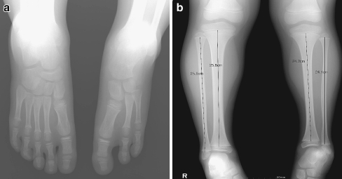
Fig. 1.
Terminal hemimelia of the lower extremity patient. a. Radiograpy of foot with two lateral rays absence. b. Proximal fibular epiphysis is at the level of the proximal tibial physis and the distal fibular physis is no higher than the tibial plafond in terminal hemimelia. The length discrepancies of fibulae between both limbs were same as those of tibiae

