
Figure 2. Raising intracellular free Ca2+ desensitizes ‘on’ bipolar cell light responses.
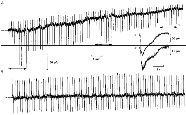
Whole-cell recordings from ‘on’ bipolar cells with 50 μm (A) and 1 μm (B) free Ca2+ in the patch pipette solution (Cs+-based) containing 5 mM BAPTA. The records begin 30 s after going whole-cell, with the cells voltage clamped to their dark potentials (-29 mV (A) and -26 mV (B)). Inward current responses were elicited by 1 Rh* test flashes (A) and 2 Rh* test flashes (B). In A, the intensity-response relation was determined at intervals (horizontal arrows) by applying light flashes which increased by factors of 2 from 0.125 to 16 Rh*. The upward deflections are current responses to 0.5 mV voltage command pulses. There was a decrease in input conductance from 43 to 14 nS as Ca2+ diffused into the cell, which was accompanied by an outward current of 35 pA from the initial dark level. The inset shows the time course of peak flash responses before (c) and after (d) equilibration with 50 μm Ca2+, as indicated.