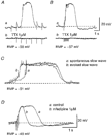
Figure 6. Effects of tetrodotoxin and nifedipine on responses produced by transmural nerve stimulation.

Aa, trains of impulses (supramaximal currents, 100 μs, 1 Hz, 5 s) initiated EJPs and triggered a slow wave at the third impulse. Ab, both EJPs and the evoked slow wave were abolished by 1 μm tetrodotoxin (TTX). Ba, trains of impulses (supramaximal currents, 100 μs, 10 Hz, 1 s) invariably evoked a slow wave which was abolished by 1 μm tetrodotoxin (Bb). An evoked slow wave (Cb) had similar amplitude and time course to a spontaneous slow wave (Ca). Da, nifedipine (1 μm) reduced the duration of the slow wave without reducing its amplitude or rise time (Db). A and B were recorded from one preparation; C and D were recorded from 2 different preparations. The scale bars in B also refer to A. The scale bars in D also refer to C. RMP, resting membrane potential.
