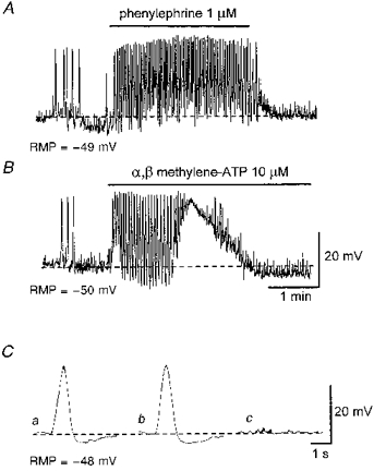
Figure 7. Effects of phenylephrine, α,β-methylene-ATP and guanethidine on slow waves.

A, phenylephrine (1 μm) increased the frequency of spontaneous slow waves and caused a sustained depolarization. B, α,β-methylene-ATP (10 μm) caused a transient increase in the frequency of spontaneous slow waves and a transient depolarization. Ca, an averaged slow wave recorded in the presence of 1 μm nifedipine. Cb, guanethidine (10 μm) did not effect the evoked slow wave. Cc, α,β-methylene-ATP (10 μm) abolished both the evoked slow wave and EJPs. Each trace in C is an average of 3-5 recordings. The scale bars in B also refer to A. The scale bars in C refer to all traces in C. RMP, resting membrane potential.
