
Figure 4. Hypoxic elevation of serotonin ([5-HT]o) levels in the extracellular fluid of the ventral respiratory region.
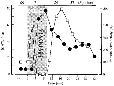
The increase in 5-HT concentration (•) coincided with secondary depression and hypoxic apnoea. The severity of hypoxia (grey area) was reduced from 6 to 10 % O2 by vol. in order to prolong the duration of the hypoxia test. Values of tissue oxygen pressure (tPO2) before, during and after hypoxia are shown. 5-HT levels remained elevated when hypoxia was terminated after 9 min. Relative changes of phrenic nerve activity (□; % of control) were calculated from its moving average.