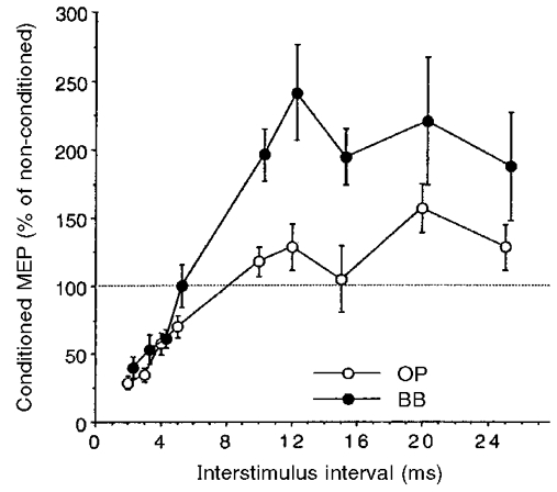
Figure 1. Intracortical inhibition and facilitation of distal and proximal arm muscles induced by paired transcranial magnetic cortical stimulation.

Time course of paired-pulse inhibition (at ISIs of 2-5 ms) and facilitation (at ISIs of 10-25 ms) in the opponens pollicis (OP; ○) and biceps brachii (BB; •) muscles. A circular coil was used and stimulus intensity was set at 80 % of the rest motor threshold (MTh) for the conditioning stimulus and at 125 % of the MTh for the test stimulus for both muscles. Each point corresponds to the mean size (±s.e.m.) of the conditioned motor evoked potential (MEP) in 9 subjects. Changes induced by the conditioning subthreshold stimuli were significantly different in the two muscles. In the BB, inhibition at ISIs of 2-5 ms was less marked and facilitation at ISIs of 10-25 ms was more marked than in the OP. Note that the filled circles are slightly shifted to the right to avoid superimposition of error bars.
