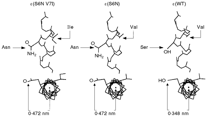
Figure 9. Longitudinal and axial views of α-helical M2 domain showing ε(S6N V7I), ε(S6N) and ε(WT) constructs.

The asparagine residue projects 0.472 nm into the lumen of the channel, as compared with 0.348 nm for serine. SwissPdbViewer (Glaxo Inc.) was used to model the helix.
