
Figure 2. BzATP induces a rise in [Ca2+]i in the paranodal Schwann cell cytoplasm.
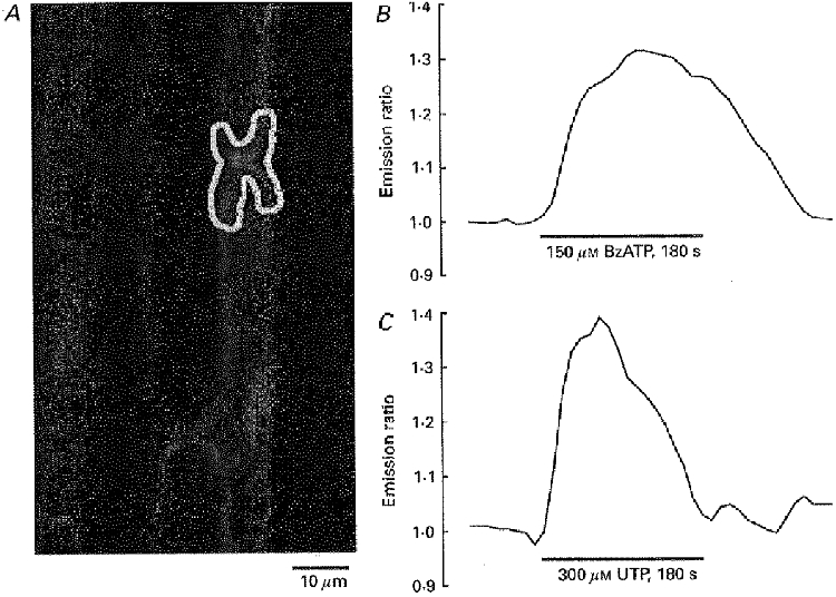
A, confocal image of myelinated nerve fibres within an isolated rat spinal root stained with the Ca2+-sensitive dyes Calcium Green-1 and Fura Red (excitation wavelength 488 nm; emission wavelengths 530 and 660 nm, respectively). B and C, the mean grey value of the paranodal Schwann cell area (selected region of interest indicated in A) and its normalized ratio was measured during bath application of BzATP (150 μM; B) and UTP (300 μM; C). Note the maintained rise in [Ca2+]i during application of BzATP whereas UTP induced a transient rise in [Ca2+]i only.