
Figure 5. Diameter distribution of DRG neurones expressing α2A-AR-IR after complete transection of the sciatic nerve 7-14 days previously.
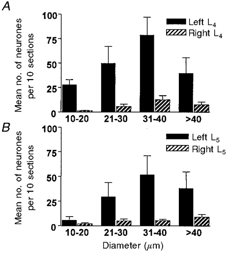
Data are means per 10 sections ±s.d. (n = 11 animals). Filled bars, ipsilateral to the lesion; hatched bars, contralateral. A, L4 DRG; B, L5 DRG.