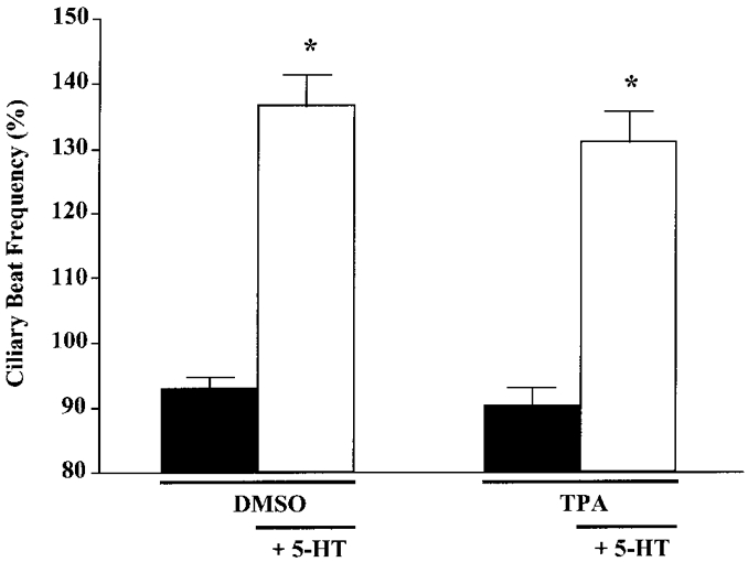
Figure 8. Effect of TPA on 5-HT-stimulated CBF.

Ciliated cells were exposed to TPA (1 μM) or DMSO (0.1%) for 10 min, followed by addition of 5-HT (100 μM) for 10 min. Addition of TPA or DMSO did not affect basal or 5-HT-stimulated CBF. Asterisks denote significant differences compared with pre-5-HT values (P < 0.05). Basal CBF values were: DMSO, 5.5 ± 0.2 beats s−1 (n = 15 cells); TPA, 5.0 ± 0.3 beats s−1 (n = 13 cells).
