
Figure 1. The stimulatory effect of adenosine is Ca2+ dependent.
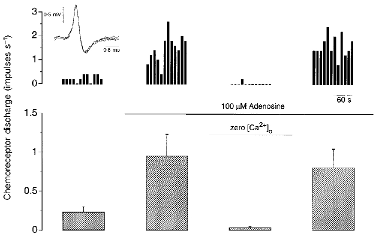
Single-fibre chemoreceptor activity recorded during steady-state control (left panel) and during exposure to 100 μM adenosine in the presence and absence of extracellular Ca2+ (middle panels) and after re-addition of Ca2+ (right panel). An example from one preparation is shown at the top, binned every 10 s and expressed in impulses s−1. The inset at top left shows three superimposed action potentials. The mean +s.e.m. of five preparations is shown below. Superfusate PO2 > 400 mmHg and PCO2≈35 mmHg throughout.