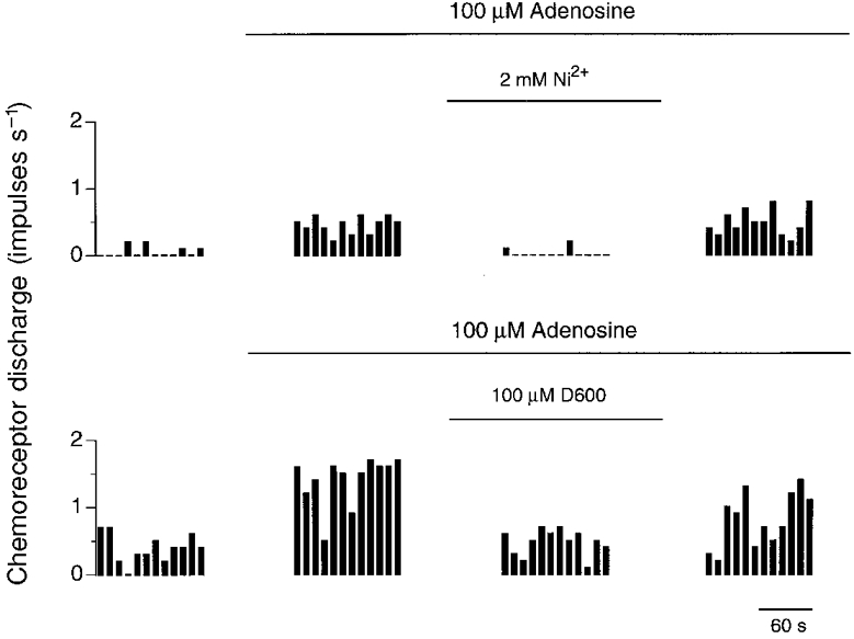
Figure 2. Effect of Ni2+ and D600 upon adenosine stimulated chemoreceptor discharge.

Single-fibre chemoreceptor activity recorded from two preparations during steady-state control (left panel) and during exposure to 100 μM adenosine in the presence and absence of either 2 mM Ni2+ or 100 μM D600 (middle panels) and after removal of antagonist (right panel). Discharge was binned every 10 s.
