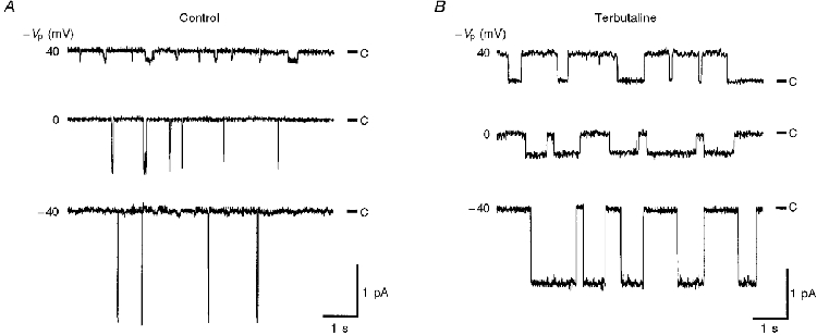
Figure 1. Single channel current traces obtained before and after application of terbutaline.

Actual traces of single channel currents obtained from a cell-attached patch formed on the apical membrane of fetal lung alveolar epithelium cultured on a permeable support before (A) and 30 min after (B) basolateral application of 10 μM terbutaline, a β2-adrenergic agonist. Voltage values (−Vp) shown next to individual traces indicate the displacement of the patch membrane potential from the resting membrane potential, i.e. 40 mV indicates that the patch membrane was depolarized by 40 mV from the membrane potential. C denotes the closed channel level. Downward deflections indicate inward current across the patch membrane (current from pipette to cell). Terbutaline activated the channel and decreased the magnitude of the inward current at −Vp= 0 mV (resting apical membrane potential). This patch membrane had only one channel.
