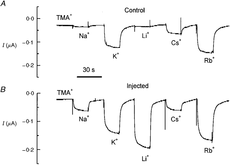
Figure 1. Ionic selectivity of KAAT1-expressing oocytes.

A, membrane currents recorded at −80 mV holding potential in a control oocyte superfused with the indicated solutions; B, same as in A for an oocyte injected with KAAT1 cRNA. Periods of 20 s in TMA+ solution separated perfusion with the various alkali ion solutions.
