
Figure 1. Video images of antral mucous cells recorded with a DIC-VEC system.
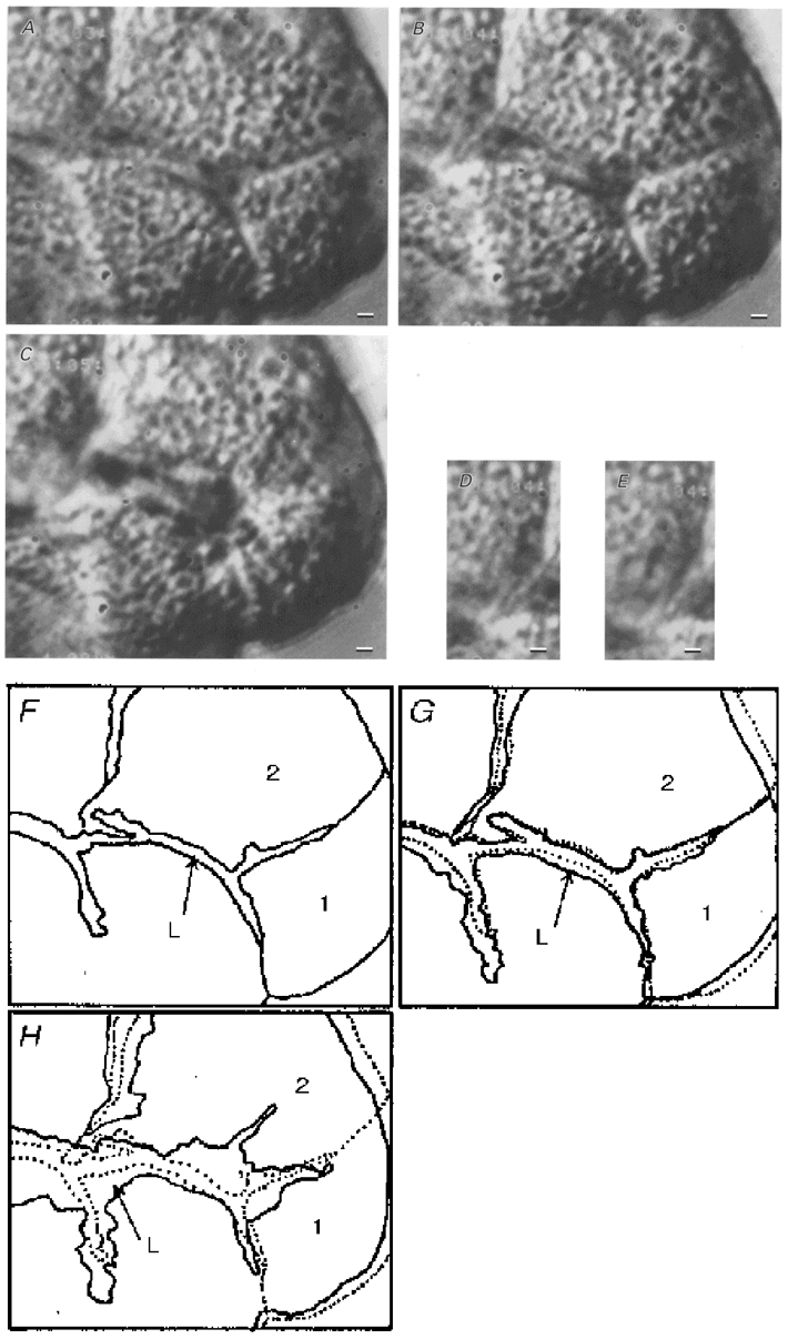
A, DIC image of unstimulated antral mucous cells densely packed with mucin granules. The lumen can be distinguished in the glandular column. B, DIC image of antral mucous cells 20 s after the start of 1 μM ACh stimulation. Cell shrinkage and swelling of the lumen are visible, but no exocytotic events were observed. C, DIC image of antral mucous cells 1 min after the start of 1 μM ACh stimulation. Exocytotic events were observed, with cells losing granules located near the luminal surface and volume at the luminal side. Swelling of the lumen increased. D and E, DIC images of mucin granules before and after exocytosis (28 and 29 s after the start of 1 μM ACh stimulation), respectively. The arrows in D show granules located near the luminal surface. After 1 s, the light intensities of the granules changed and Ω-shaped holes were observed in the luminal surface (arrows in E). F-H, outlines of the video images shown in A-C. ‘L’ is the lumen; ‘1′ and ‘2′ indicate mucous cells. The dotted lines in G and H give the outlines of the unstimulated mucous cells shown in A and F. G shows the initial cell shrinkage of antral mucous cells. H also shows the delayed cell shrinkage, especially at the luminal side, caused by granule release (exocytotic events). Scale bars represent 2 μm.