
Figure 11. Changes in [Ca2+]i in antral mucous cells.
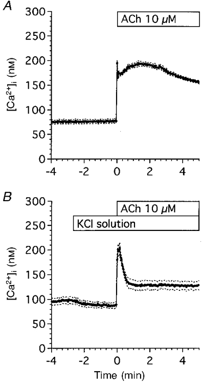
Cells were loaded with fura-2 AM for 25 min at room temperature. The ratios (340 nm/380 nm) of fura-2 fluorescence were calculated and stored in the Ca2+ imaging system. A, control experiments (n = 6). Cells were perfused with the control solution and stimulated with 10 μM ACh. ACh stimulation evoked an initial followed by a sustained increase in [Ca2+]i. B, KCl experiments (n = 5). Fura-2 AM-loaded cells were perfused with the KCl solution and stimulated with 10 μM ACh. ACh stimulation evoked an initial followed by a sustained increase in [Ca2+]i. [Ca2+]i in the sustained phase of control experiments was higher than that in KCl experiments, but [Ca2+]i during the first 30 s of ACh stimulation increased significantly in both experiments. Values are expressed as means ±s.e.m.