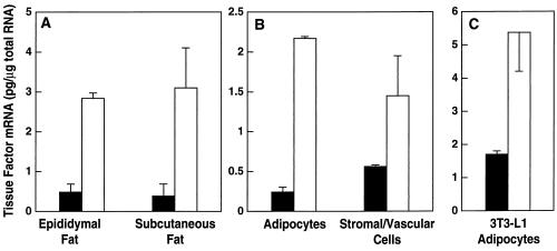
Figure 4.
Induction of TF mRNA expression in adipose tissue and adipocytes by TGF-β. (A) Six to 8-week-old male lean CB6 mice were injected i.p. with 2 μg of human recombinant TGF-β (□) or diluent (▪), and 3 h later, the adipose tissues were removed and analyzed for TFmRNA. Total RNA was prepared and analyzed for TF gene expression by quantitative RT-PCR (n = 3 ± SD). Comparison of TF mRNA levels in epididymal fat from control vs. TGF-β-treated mice using the unpaired Student’s t test reveal P < 0.01; for subcutaneous fat from control vs. TGF-β-treated mice, P < 0.01. (B) Mature adipocytes and stromal vascular cells were separated by differential centrifugation, total RNA was prepared, and TF mRNA levels were determined (n = 3 ± SD). Comparison of TF mRNA levels in adipocytes from control and TGF-β-treated mice reveal P < 0.0006; for control vs. TGF-β-treated stromal vascular cells, P < 0.1. (C) 3T3-L1 adipocytes were grown and differentiated in 6-well tissue culture plates as described (40). Total RNA was isolated from untreated cells (▪), and cells treated with 1 ng/ml TGF-β (□) for 3 h, and steady-state levels of TF mRNA were determined by using quantitative RT-PCR (n = 6 ± SD).

