
Figure 4. Representative SDS-polyacrylamide gels illustrating MHC and MLC expression in single human soleus fibres.
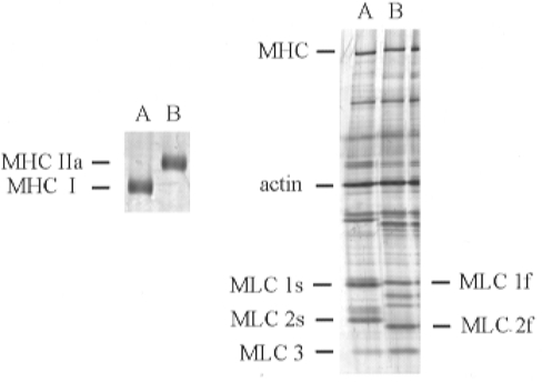
Left: 5 % gel demonstrating separation of type I and IIa MHC in two single soleus fibres. Right: 12 % gel illustrating MLC composition of the same two single fibres. Fibre Vo was 1.09 FL s−1 for the type I fibre in lane A and 4.09 FL s−1 for the type IIa fibre in lane B.