
Figure 1. Immunocytochemical characterization of the rat aortic smooth muscle cell line.
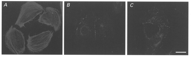
Photomicrographs showing abundance and distribution of immunoreactivity to anti-smooth muscle actin (A), anti-Cx40 (B) and anti-Cx43 (C) antibodies in A7r5 cells grown overnight under serum-free conditions. Bar, 15 μm.