
Figure 7. CaMKII-like immunoreactivity in mouse proximal colon.
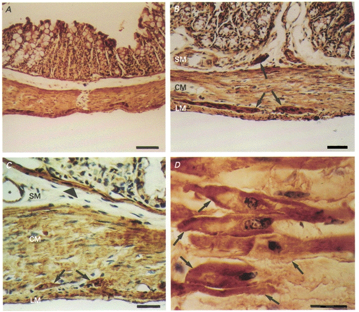
Haematoxylin counterstain. A, CaMKII-like immunoreactivity (in brown) exhibited throughout the entire external muscularis (scale bar = 100 μm). B, CaMKII-like immunoreactivity (in brown) within both longitudinal and circular muscle layers. Also, more intense staining of enteric ganglia within the deep muscular plexus of the submucosa and in the myenteric plexus between the longitudinal and circular muscle layers (arrows) (scale bar = 50 μm). C, CaMKII-like immunoreactivity (in brown) again showing positive staining of the longitudinal and circular muscle layer and enteric ganglia. Also, positive reactivity with the muscularis mucosa (arrowhead) and blood vessels (*) (scale bar = 20 μm). D, CaMKII-like immunoreactivity (in brown) of individual smooth muscle cells of the circular muscularis (arrows). Staining appears more intense at the level of the cell membrane (scale bar = 20 μm). SM, submucosa; CM, circular muscularis; LM, longitudinal muscularis.