
Figure 1. Recording of a single motor unit during finger movement.
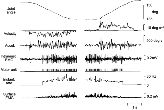
A sample extension-flexion movement trial. The target used for visual tracking moved at a fixed rate of 10 deg s−1. Records show, from above: angle at the metacarpo-phalangeal joint; angular velocity; acceleration; intramuscular EMG; a symbolic representation of the motor unit discharge train; instantaneous frequency of motor unit discharge; and the surface EMG. EMG signals were obtained from the common finger extensor muscle (m. extensor digitorum communis, EDC).