
Figure 1. Lack of activation of KACh channels by inositol-containing phospholipids and PE in inside-out patches.
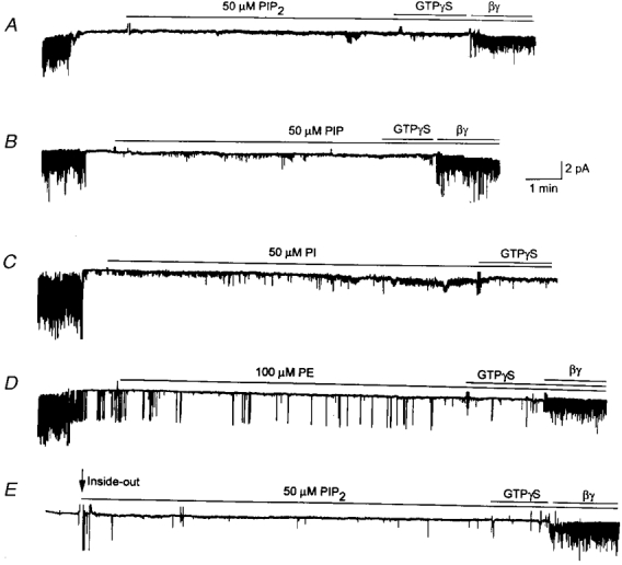
A-D, cell-attached patches were formed with 10 μM ACh in the pipette. Channels were activated when ACh was present in the pipette. Upon formation of inside-out patches, channels closed immediately. PIP2 (50 μM), PIP (50 μM), PI (50 μM) or PE (100 μM) was then applied to the cytoplasmic side of the membrane for ≈10 min. GTPγS (10 μM) was then applied for ≈1 min, and then purified bovine βγ subunit (50 nM) applied subsequently. Although not shown in C, the βγ subunit activated the channels when applied ≈5 min after the GTPγS application. In D, occasional openings of one KATP channel can be seen. In E, no ACh was added to the pipette solution. Membrane potential was held at -60 mV. All tracings were filtered at 100 Hz.