
Figure 2. Intracellular CO2-dependent buffering.
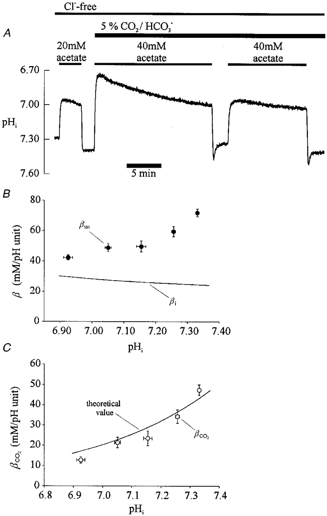
A, intracellular buffering doubles on switching from Hepes to CO2/HCO3−-buffered conditions. The trace shows a ratiometric recording of pHi. Superfusates were Cl− free (gluconate substituted). An acetate prepulse (20 mM) was performed initially in Hepes, then (40 mM) in CO2/HCO3−-buffered solutions (same pHo= 7.40). B, total intracellular buffering power (βtot) in CO2/HCO3−-buffered conditions. Experimental protocol: a 40 mM acetate prepulse was executed in Cl−-free, 5 % CO2/HCO3−-buffered solution (pHo 7.40) and the subsequent steady-state rise of pHi used to calculate intracellular buffering power (βtot). This has been plotted versus pHi at the mid-point of the rise of intracellular HCO3− following acetate removal (see text for details). Data have been pooled from several cells and averaged for the following 0.1 pHi ranges (•) starting at pH 6.90, with sample sizes, n = 8, 9, 6, 19 and 20. Also plotted (lower continuous line) is the relationship between intrinsic buffering capacity (βi) and pHi over the same pHi range (6.90-7.38) calculated from eqn (1) in Results. C,βCO2 in the cardiac cell. At a given pHi, the difference between the mean experimental values of βtot (• in B) and the value of βi determined from eqn (1) (continuous curve in B) gives βCO2. This difference has been plotted versus pHi in C (○). The continuous line drawn through the data is the theoretical value of βCO2vs. pHi for a cell open to CO2, and has been calculated from eqn (2) in Results.