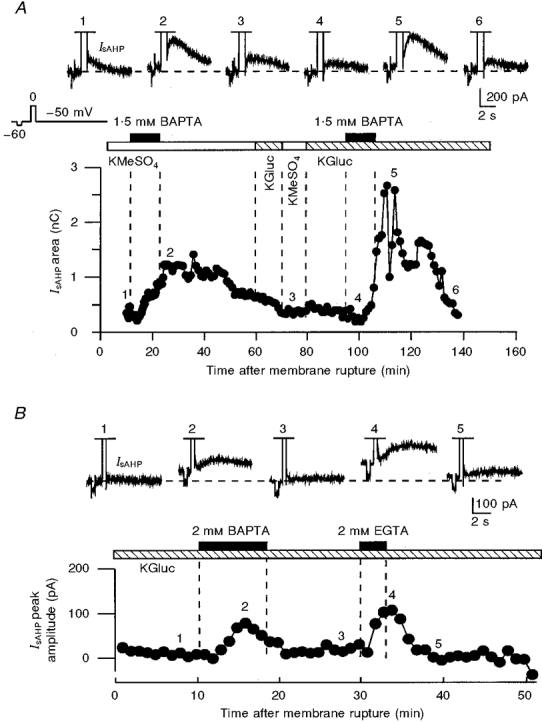
Figure 1. Potentiation of IsAHP by internally perfused low millimolar concentrations of BAPTA and EGTA in two voltage-clamped neurones.

A, the potentiating effects of BAPTA could be reproduced in the same neurone with both methyl sulphate- and gluconate-based internal solutions. Initially, 1.5 mM BAPTA was applied in the presence of a KMeSO4-based internal solution. During the second hour of recording, after washout of BAPTA, the internal solution was replaced with a potassium gluconate-based solution (KGluc), and 1.5 mM BAPTA was applied again. B, effects of internally perfused 2 mM BAPTA and 2 mM EGTA in the presence of potassium gluconate-based internal solution in another neurone. In B, the IsAHP amplitude (measured from the holding current level), but not the area, was plotted in the graph because most of the current was off the recorded time frame. The slower effects of internal perfusion in A were due to a higher access resistance (A, 15 MΩ; B, 6 MΩ). In A and B, the numbers above the traces correspond to the numbers on the graphs. A -10 mV voltage step from a holding voltage of -50 mV, preceding the depolarizing voltage command used for activating Ca2+ influx into the cells, was used for monitoring the resting membrane conductance.
