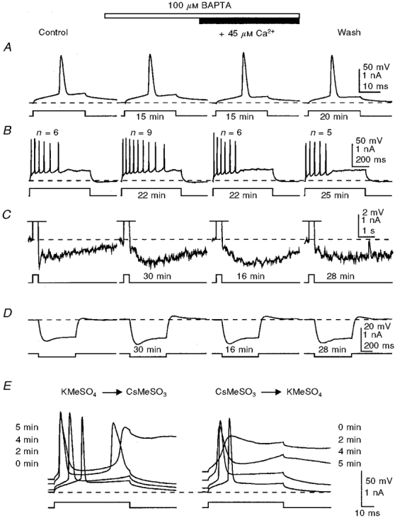
Figure 3. Addition of Ca2+ to restore the control [Ca2+]i does not reverse the effects of submillimolar concentrations of BAPTA on the mAHP/sAHP, but does reverse the effect on spike firing pattern.

The mAHP/sAHP (C) was activated by trains of action potentials (shown in B; off scale in C) generated during depolarizing current injections. Note the absence of BAPTA effects on the action potential (A) and the reduction of the mAHP during potentiation of the sAHP (C). At concentrations of 100-300 μM, BAPTA caused a slight increase in the membrane input resistance, as revealed by voltage responses to hyperpolarizing current steps (D). E, the effectiveness of internal perfusion was tested in this cell after washout of BAPTA by replacing internal K+ with Cs+ (see Methods). Note the recovery of the number of action potentials (n) generated by a depolarizing pulse after addition of Ca2+ to the BAPTA-containing solution (B), and a correspondence between these changes and the onset of the sAHP. The times shown indicate the duration of perfusion of a particular solution before the traces were taken. The membrane potential was held at -60 mV (dashed lines). KMeSO4-based internal solution.
