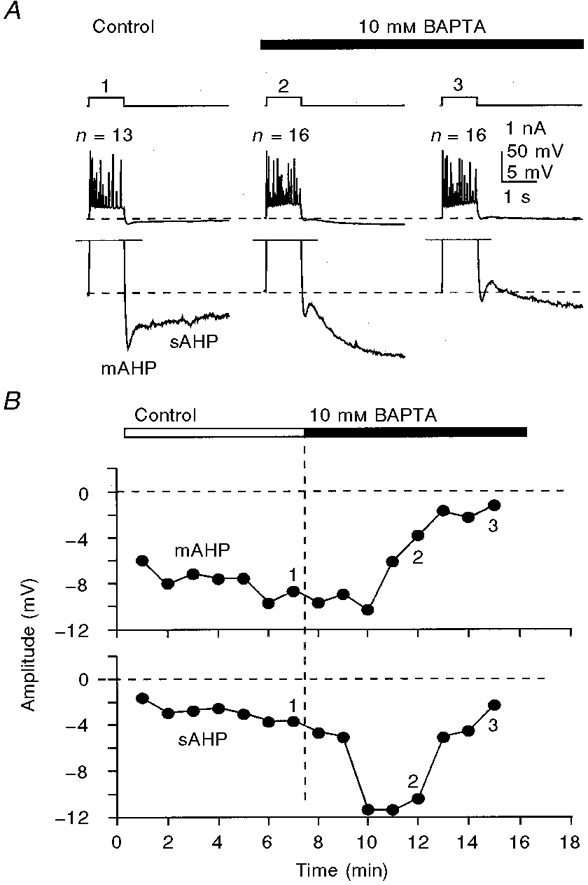
Figure 5. Transient potentiation of the sAHP and prolonged inhibition of the mAHP by internally perfused 10 mM BAPTA.

A, sample responses to 400 pA, 800 ms depolarizing current pulses taken at corrresponding times shown by numbers on the graph (B). Note the differences in the time courses of the inhibition of the mAHP and potentiation of the sAHP during BAPTA perfusion. The appearance of a small depolarizing component separating the mAHP and the sAHP at later stages of BAPTA perfusion (A3) was a common phenomenon with 10-15 mM concentrations of BAPTA. The membrane potential was held at -62 mV (dashed lines in A). The number of action potentials (n) generated by depolarizing pulses is shown above the traces in A. KMeSO4-based internal solution.
