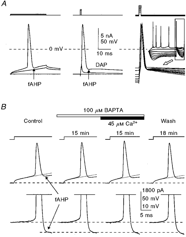
Figure 8. Submillimolar concentrations of BAPTA do not affect the size and shape of action potentials and the amplitude of the fAHP both without and with added Ca2+.

A, comparison of fAHPs following action potentials activated by long (left) and short (middle and right) depolarizing current pulses. B, separation of the fAHP (recorded in the cell shown in Fig. 3) by digital subtraction of the normalized subthreshold voltage transient (shown with dotted lines on upper traces) from the traces with action potentials, the results of which are shown in the lower traces. The times given indicate the duration of internal perfusion of the particular solutions indicated. The membrane potential was held at -60 mV (dashed lines). KMeSO4-based internal solution.
