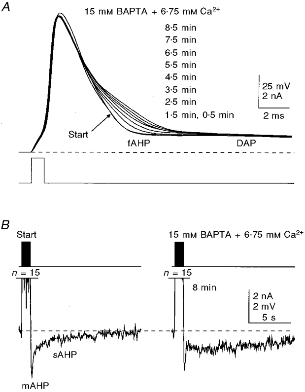
Figure 9. Blockade of the fAHP and prolongation of the action potential decay with Ca2+-balanced 15 mM BAPTA.

In this cell, the whole-cell recording was started with a patch pipette loaded with 15 mM BAPTA. A, prolongation of the second half of the decay of the action potential caused by BAPTA. B, in the same cell, the mAHP/sAHP showed changes that were qualitatively similar to those observed at lower BAPTA concentrations. Inhibition of the mAHP seen in B began before the trace marked Start was recorded (0.5 min after establishing the whole-cell recording configuration) due to the possible faster diffusion of BAPTA into the cell caused by a very high concentration gradient. The membrane potential was held at -62 mV (dashed lines). Potassium gluconate-based internal solution. n, number of action potentials. The times shown indicate the duration of internal perfusion of the BAPTA solution before the traces were taken.
