
Figure 1. Current-voltage relations of peak outward K+ currents in isolated adult and postnatal day 28 (P28) rat atrial myocytes are indistinguishable.
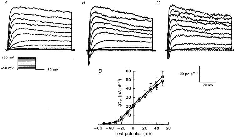
Outward currents, evoked during 100 ms depolarizing voltage steps to potentials from -50 to +50 mV from a holding potential of -60 mV, were recorded in individual cells and normalized to the whole-cell membrane capacitance, Cm (determined in the same cell). Representative current waveforms, normalized for difference in cell sizes, recorded in adult (A) and in postnatal day 28 rod-shaped (P28R) (B) and spherical (P28S) (C) rat atrial myocytes are displayed. D, mean ±s.e.m. peak outward current density for the currents recorded in P28S (n = 25), P28R (n = 9) and adult (n = 11) cells are plotted as a function of test potential. One-way analysis of variance (ANOVA) revealed no significant differences in peak current-voltage relations in adult (○), P28S (⋄) and P28R (□) atrial myocytes.