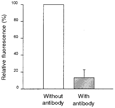
Figure 4. Quenching of the fluorescence intensity of a dye-labelled epithelium by the addition of a monoclonal antibody against free and bound fluorescein.

The fluorescence intensity (λEx 436 nm) was measured directly after staining the epithelium with HAF and 45 min after addition of the monoclonal IgG antibodies against fluorescein (shaded bar) (n = 3). Results are given as percentage of the dye-loaded epithelium.
