
Figure 1. Distribution of neurones immunoreactive for both PNMT and TH (C1 cells) and neurones containing only TH immunoreactivity (A1 cells).
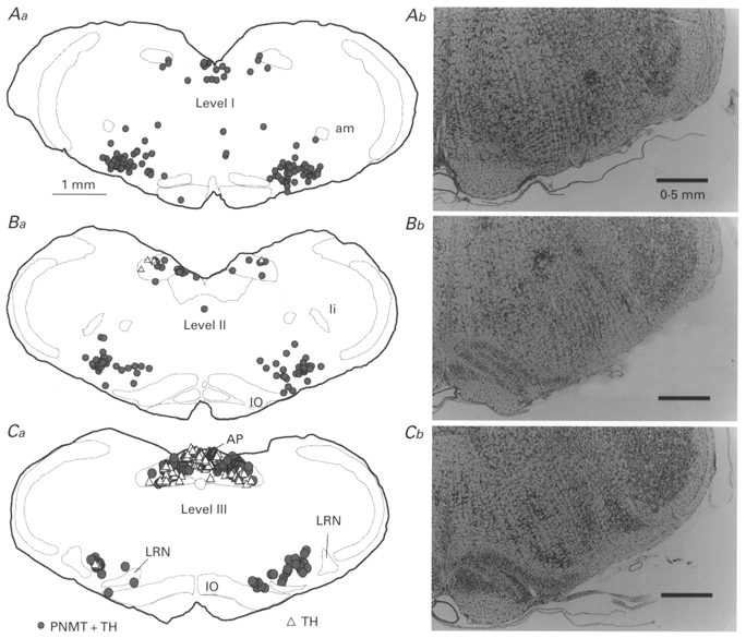
Distribution is shown at the three rostrocaudal levels where the electrophysiological recordings were made. Level I is just caudal to the facial motor nucleus (Aa; 11.8 mm caudal to bregma, co-ordinate after Paxinos & Watson, 1986), level II at an intermediate rostrocaudal location (Ba; 12.5 mm caudal to bregma), and level III at the level of the rostral border of the lateral reticular nucleus (Ca; 13.1 mm caudal to bregma). Corresponding Nissl-stained sections are shown for comparison (Ab-Cb). Abbreviations: am, nucleus ambiguus, compact portion; AP, area postrema; IO, inferior olive; li, nucleus linearis; LRN, lateral reticular nucleus.