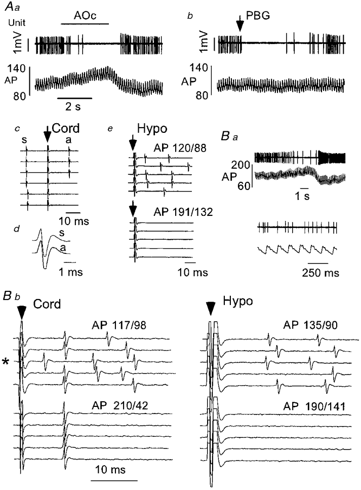
Figure 3. Physiological properties of a type I phenotypically identified C1 neurone.

Aa, elevation of arterial blood pressure (AP, mmHg) by gradual aortic occlusion (AOc) resulted in inhibition of the discharge of this cell. Ab, cardiopulmonary receptor stimulation (PBG, 10 μg kg−1, i.v.) briefly silenced the discharge of this neurone. Ac, constant latency antidromic spike (a) elicited by spinal cord stimulation (arrow; traces 1-3 from top). The delay between the spontaneous spike (s) and the spinal stimulus exceeds the critical interval for collision. Reduction of the delay between the spontaneous spike and the spinal stimulus to the critical interval results in collision of the antidromic spike (traces 4-6). Ad, configurations of the spontaneous (s) and antidromic spikes (a). Ae, hypothalamic stimulation (arrow) at resting arterial blood pressure (AP: 120/88 mmHg) results in orthodromic activation of the cell that is abolished by AP elevation (aortic occlusion). Note the absence of constant latency spikes. Ba, a second example of a type I barosensitive cell. The discharge of the cell was reduced during arterial blood pressure elevation (aortic occlusion). The discharge of the cell at resting arterial blood pressure is also shown on an expanded time scale. Bb, spinal cord stimulation (left panel) at resting arterial blood pressure elicits constant latency (antidromic) spikes (traces 1, 2, 4 and 5) as well as orthodromic responses. Collision of the antidromic spike with a spontaneous spike is evident in trace 3 (marked with an asterisk). Elevation of arterial blood pressure eliminates orthodromic driving but not antidromic activation. Hypothalamic stimulation (right panel) elicits only orthodromic spikes which are eliminated by arterial blood pressure elevation. No constant latency spikes are evident.
