
Figure 5. Physiological properties of a type II phenotypically identified C1 neurone recorded in a rat ventilated with room air.
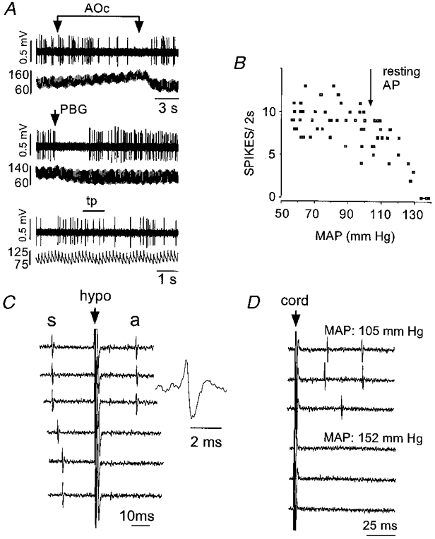
A, inhibition by aortic occlusion (AOc) (top two traces), i.v. injection of phenylbiguanide (PBG, 10 μg kg−1, middle pair of traces) and insensitivity to nociceptive stimulation (toe pinch, tp; bottom pair of traces). In each pair of traces, the top trace shows the unit activity and the bottom trace AP (mmHg). B, relationship between discharge rate and mean arterial blood pressure. C, antidromic activation by hypothalamic stimulation. Hypothalamic stimulation (at arrow) triggered by the occurrence of a spontaneous spike (s) elicits constant latency antidromic spikes (a; top three traces). Stimulation within the critical latency results in collision of the antidromic spike (bottom three traces). The inset shows an enlargement of the antidromic spike. D, orthodromic driving (top three traces) produced by stimulation of the spinal cord (at arrow) was abolished by elevation of arterial blood pressure (bottom three traces). Peak to peak amplitude of spikes in C and D: 0.8 mV.