
Figure 11. Phenotypic characterization of VLM neurones.
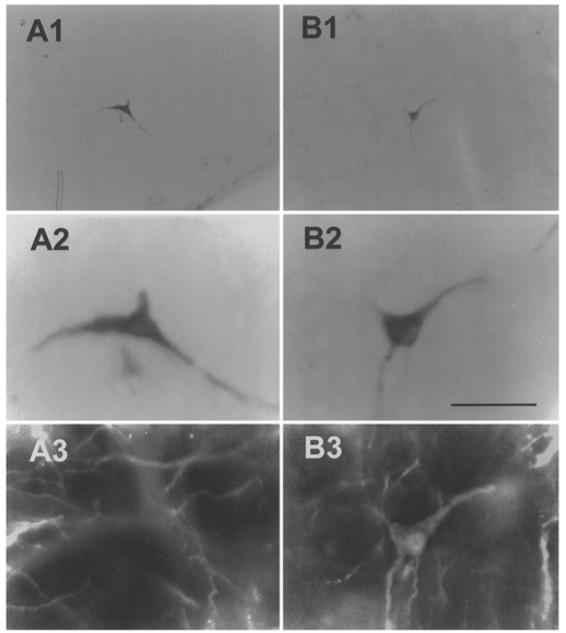
A1, low power photomicrograph of a single biotinamide-labelled ventrolateral medullary barosensitive, medullospinal neurone (type I). A2, high power photomicrograph of cell in A1. The dark material immediately below this neuron was an artifact associated with a small amount of damage caused by the microelectrode. A3, same cell is devoid of PNMT immunofluorescence. B1, low power photomicrograph of a single biotinamide-labelled ventrolateral medullary barosensitive neurone projecting to the hypothalamus (type II). B2, high power photomicrograph of cell in B1. B3, same cell as in B1 and B2 displaying immunofluorescence for PNMT. Scale bar for A2, A3, B2 and B3 is 50 μm; scale bar for A1 and B1 is 200 μm.