
Figure 12. Location, phenotype and projection pattern of histologically recovered neurones.
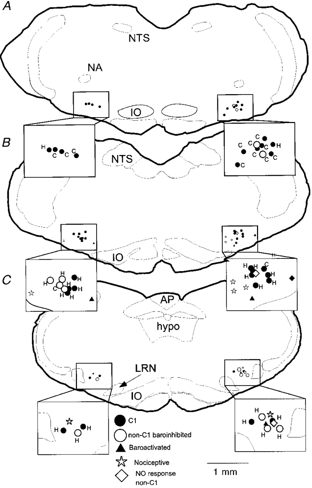
A, B and C depict neurones identified in levels I, II and III, respectively. •, C1 cells (includes types I, II and III); ○, non-C1 barosensitive cells (types I, II and III); ▴, baroactivated cells (type VI); *, nociceptive cells (type IV); ⋄, non-responsive, non-C1 cells (type V). Each inset shows an enlargement of the VLM area and identifies whether the baroinhibited cells have spinal (C) or hypothalamic (H) projections. Abbreviations: AP, area postrema; hypo, hypoglossal nucleus; IO, inferior olive; LRN, lateral reticular nucleus; NA, ambiguus nucleus; NTS, solitary tract nucleus.