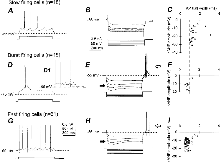
Figure 2. Identification of slow firing, burst firing and fast firing neurons in the medial septum-diagonal band.

A, D and G, responses to injection of depolarizing current pulses, as indicated by the square pulses beneath the traces. B, E and H, overlay of responses to a series of hyperpolarizing current pulses, indicated beneath the traces. C, F and I, scatter plots for all cells showing relationship of action potential (AP) half-width with slow after-hyperpolarization (sAHP) amplitude. Slow firing neurons fire slow repetitive action potentials followed by pronounced, long-lasting after-hyperpolarizations (A) and lack time-dependent inward rectification, or depolarizing sag (B). They can be clearly distinguished from fast firing and burst firing neurons on the basis of their action potential waveform (C, F and I). Burst firing neurons show a burst of action potentials overriding a slow depolarization in response to a threshold depolarizing current pulse from −75 mV and repetitive firing when depolarized from - 55 mV (D and D1), and have depolarizing sag (filled arrow in E) and rebound firing (open arrow in E). Fast firing neurons possess higher firing rates and smaller after-hyperpolarizations (G) than slow firing neurons, and show depolarizing sag (filled arrow in H) and rebound firing (open arrow in H).
