
Figure 2. Simultaneous localization of immunoreactive PAR-1 and PAR-2 with PGP 9.5, SP, NOS and VIP in myenteric neurons in culture.
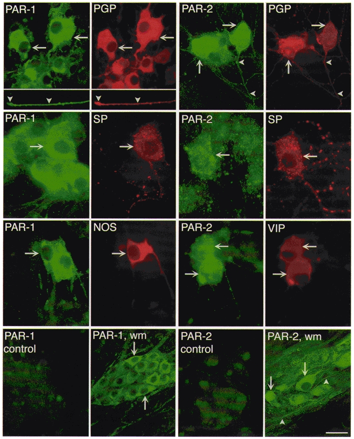
To localize PAR-1, cultures were incubated with mouse PAR-1 61-1 and rabbit PGP 9.5, or rat SP, or rabbit NOS antibodies. To localize PAR-2, cultures were incubated with rabbit PAR-2 B5 and mouse PGP 9.5 antibodies, or rabbit PAR-2 9717 and rat SP, or mouse VIP antibodies. The arrows indicate colocalization in neurons of PAR-1 and PAR-2 with PGP 9.5, SP, VIP or NOS. The arrowheads indicate stained neurites. In the control experiments, antibodies to 61-1 or B5 were preincubated with 10 μm of the peptides used for immunization. PAR-1 wm (whole mount) and PAR-2 wm show localization in whole mounts of the myenteric plexus using 61-1 and 9717, respectively. Scale bar, 20 μm for top row and controls, 28 μm for middle two rows, and 50 μm for whole mounts.