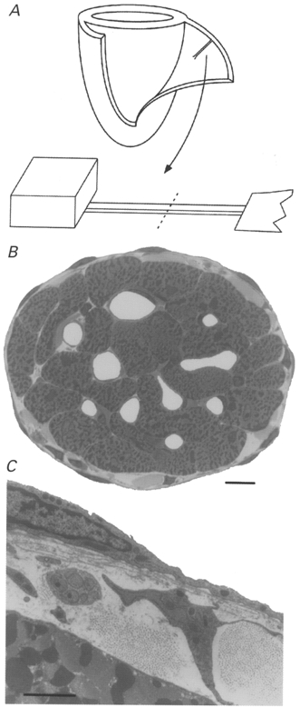
Figure 1. Ultrastructure of rat cardiac trabeculae.

A, schematic diagram showing a suitable trabecula before and after its isolation from the right ventricular free wall of a glutaraldehyde-fixed heart (only ventricles depicted). The dashed line indicates where the trabecula was transversely sectioned after resin embedding. B, low-power image, obtained by light microscopy, of the cross-section of a trabecula. Scale bar, 10 μm. C, electron micrograph of the periphery of the above trabecula. Note the presence of a fibroblast, a bundle of nerve axons and fibrillar collagen in the space between the endothelium and a myocyte. Scale bar, 2 μm.
