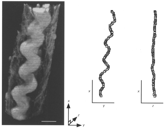
Figure 3. 3-D reconstruction of a typical perimysial collagen fibre in a ventricular trabecula fixed at near-resting sarcomere length.

The trabecula was fixed at a sarcomere length of 1.81 μm and stained with a collagen-specific fluorophore (Sirius Red F3BA). Confocal laser scanning microscopy (via a × 100 objective lens) was used to obtain the 30 serial optical images (taken at 0.39 μm intervals) used in this reconstruction. 3-D analysis of this fibre showed that it was wavy in a planar fashion rather than coiled. White scale bar, 10 μm. On the right, the centre of the fibre has been plotted in orthogonal planes. Note that the fibre is wavy in the X-Y plane (viewed from above) whereas it is not in the orthogonal X-Z (or profile) view. The axes serve as scale bars and are each 20 μm.
