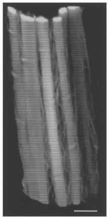
Figure 4. 3-D reconstruction of perimysial collagen fibres in a ventricular trabecula fixed at an extended sarcomere length.

The preparation was fixed at a sarcomere length of 2.27 μm. Confocal laser scanning microscopy (via a × 63 objective lens) was used to obtain the fifty optical sections (taken at 0.31 μm intervals) used in this reconstruction. Note that some of the fibres show small kinks. Scale bar, 10 μm.
