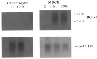
Figure 2. Failure to detect mRNA for BGT-1 in chondrocytes.

MDCK cells and primary cultures of chondrocytes were incubated, separately, in control medium (C, 0.3 osmol l−1) or test medium (T, 0.5 osmol l−1) for the indicated periods of time. Poly(A)+RNA was then extracted from the cells and analysed by Northern blotting for the presence of BGT-1 mRNA and β-actin mRNA, as described in the Methods section. The blot of β-actin mRNA was used to monitor the uniformity of loading the gel with the different samples of poly(A)+RNA: clearly, there were no significant differences between control and test samples in chondrocytes.
