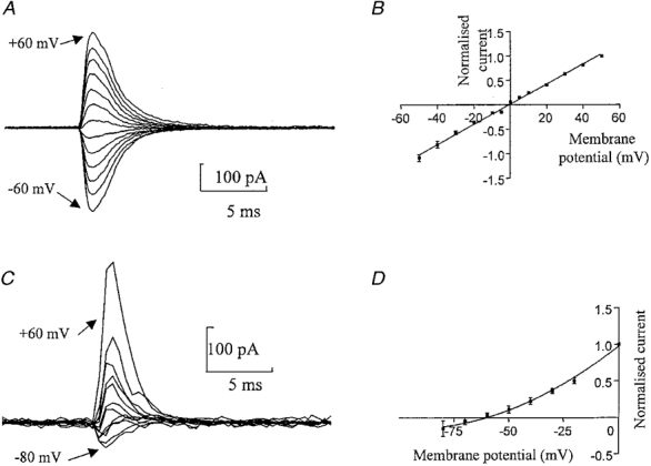
Figure 2. Current-voltage relationship and calcium permeability of somatic GluR channels.

A, currents from a nucleated patch activated by 2 ms pulses of L-glutamate (1 mM) in normal Hepes-based solution. In this cell the responses were measured at different membrane potentials ranging from -60 to +60 mV in 10 mV steps. Each trace is an average of at least three responses. B, normalized current- voltage relationship of the peak glutamate-activated current for 4 patches. The data were fitted by a straight line giving an estimate for the reversal potential of -0·6 mV (corrected for liquid junction potential). C, currents from a nucleated patch activated by a 2 ms pulse of L-glutamate (1 mM) recorded in a Ca2+-rich extracellular solution, using a Cs+-rich intracellular solution. The membrane potential was held at 60, 40, 20, 0, -10, -20, -30, -40, -50, -60, -70 and -80 mV. Currents were digitized at 0·5 ms. D, normalized current-voltage relationship for all 6 patches. The data were fitted by a second order polynomial (r2= 0·9994), giving a reversal potential of -63·25 mV (n= 6) (corrected for liquid junction potential). Currents were normalized to +50 mV in B and 0 mV in D.
