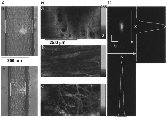
Figure 1. Optical sectioning of the vascular wall of pressurized mesenteric arteries and determination of optical performance in situ.

A, transmitted light images of a pressurized (70 mmHg) rat mesenteric artery mounted in a recording chamber on the microscope when fully relaxed (diameter, 224 μm) in Ca2+-free-2 mM EGTA Krebs solution (a) and after developing myogenic tone (136 μm) in normal Krebs solution (b). White bars are the video calipers used to measure lumen diameter. B, high-resolution optical sections of an artery loaded with Calcium Green-1 showing that discrete regions of the arterial wall can be visualized. The lumenal endothelium (a), media with smooth muscle cells (b), and perivascular neurons (c) are readily distinguished. C, point-spread function (PSF) of the microscope obtained by imaging a single fluorescent bead (0.1 μm diameter) that had been made to lodge on the inner surface of a typical arteriole not itself loaded with fluorescent dye. The x-z plane image was obtained by slicing vertically a 3-dimensional reconstruction of the bead obtained with pixels of 0.1 μm in the x-, y- and z-axes. The shifts of successive x-y planes apparent in the bead image were due to slight movement of the artery. These shifts, of approximately 0.1 μm, were not observed with beads adhering to the coverslips.
