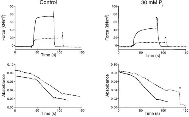
Figure 1. Force and ATPase activity in a mechanically skinned bundle of myofibrils in the absence and presence of 30 mM Pi.

The dashed lines represent results obtained in the presence of 10 mM BDM. Upper recordings, force development; lower recordings, NADH absorbance. The preparation was activated by transferring it from the pre-activating solution (pCa 9) into the activating solution (pCa 4.8), and relaxed at the end of the measurement by transferring it back into relaxing solution (pCa 9). The final slope of the absorbance signal, relative to absorbance baseline (measured when the preparation was not in the measuring chamber), was used as a measure of the rate of ATP consumption. At the asterisk (lower right panel) a calibration of the absorbance signal is shown which corresponded to 0.5 nmol of ATP hydrolysed. The zero level in the absorbance signal was arbitrarily chosen. Preparation diameters (measured in two perpendicular directions), 160/140 μm; preparation length, 1.85 mm.
