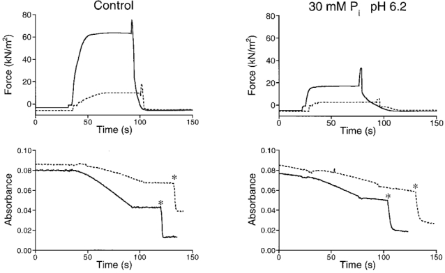
Figure 5. Combined effects of 30 mM Pi and pH 6.2 on force and ATPase activity in a mechanically skinned bundle of myofibrils.

The results at pH 7.1 (left) are shown in the absence and presence (dashed lines) of 10 mM BDM. In the right panels, the corresponding results are shown obtained at 30 mM Pi and pH 6.2. Upper recordings, force development; lower recordings, NADH absorbance. The slower responses upon calibration injections (∗) in the right recordings, as compared with the left recordings, reflect the reduction in PK activity at pH 6.2. Preparation diameters, 140/130 μm; preparation length, 3.50 mm.
