
Figure 2. Activation of ionic currents by IP3, IP4 and thapsigargin in CHO(CCE1) cells.
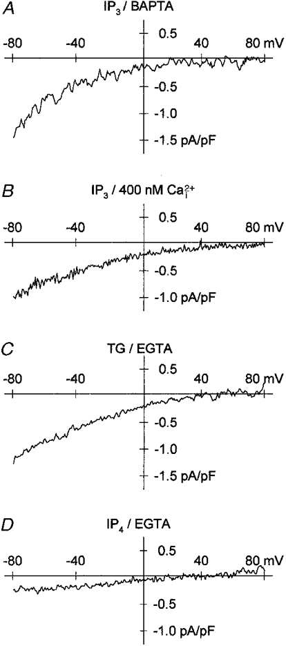
The internal solution contained 115 mM Cs+. IP3 (10 μM) was dialysed in the presence of 10 mM BAPTA (A, IP3/BAPTA) and 400 nM free Ca2+ (B, IP3/400 nM ). Thapsigargin (1 μM) was applied to cells dialysed with 10 mM EGTA (C, TG/EGTA). IP4 (20 μM) was dialysed with 10 mM EGTA (D, IP4/EGTA). Same external solution as in Fig. 1.