
Figure 7. Removal of external Ca2+ or Mg2+ does not affect capsaicin-induced inward current after prior desensitization of the response to capsaicin.
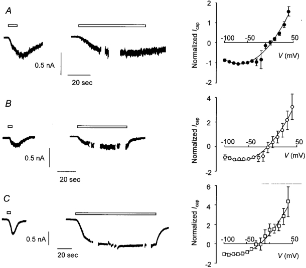
A, trace from a voltage-clamped DRG neurone exposed to Ca2+ (1 mM)- and Mg2+ (1 mM)-containing external solution throughout. Capsaicin (0.5 μM) was applied as a conditioning pulse for 5 s to produce desensitization, then re-applied as a test pulse for approximately 60 s during which time the I-V curve was determined. The right-hand panel shows the I-V relationship for capsaicin-activated current obtained during the test pulse (means ±s.e.m., n = 5). B, current recorded from a second DRG neurone. Data were obtained using the same protocol as in A except that the test pulse was applied in the absence of Ca2+ (no added Ca2+, 100 μM EGTA). The right-hand panel shows the I-V relationship obtained during the test pulse (means ±s.e.m., n = 8). C, trace obtained from a third DRG neurone. The protocol was the same as in A and B except that the test pulse (120 s) was applied in the absence of Ca2+ and Mg2+. The right-hand panel shows the I-V relationship obtained during the test pulse (means ±s.e.m., n = 5). For each trace open bars indicate application of capsaicin and breaks in the trace during capsaicin application show the points at which I-V curves were generated. Vh was -60 mV. The points on each I-V curve are joined by a polynomial curve fitted after omitting data from potentials between -20 and +10 mV although all data points have been plotted. Background current was subtracted and current was normalized to the inward current at the Vh of -60 mV.