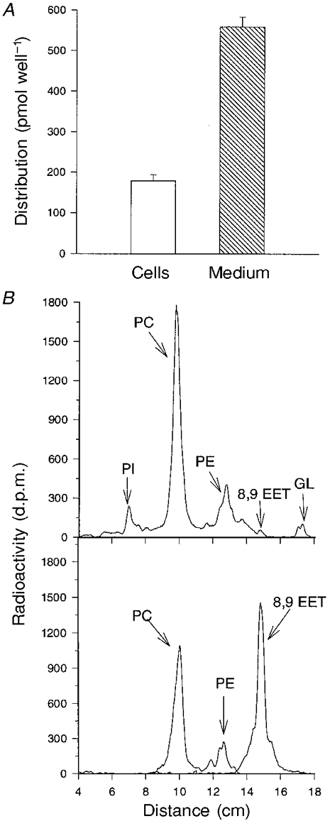
Figure 9. Distribution of radioactivity following incubation of rat neonatal myocytes with [3H]8,9-EET.

Cells were incubated in medium containing 1 μM [3H]8,9-EET for 30 min to 4 h, after which the cell- and medium-associated lipids were extracted and analysed. The distribution of radioactivity between the cells and medium (quantified by liquid scintillation counting) following a 2 h incubation with [3H]8,9-EET is shown in A. The values are expressed as means ± s.e.m., n = 3. In B, a representative TLC chromatogram (upper panel) shows the distribution of radioactivity in cell lipids following a 30 min incubation with [3H]8,9-EET. PI, phosphatidylinositol; PC, phosphatidylcholine; PE, phosphatidylethanolamine; GL, glycerides. Similar results were obtained from a duplicate culture. Migration of radiolabelled phospholipid standards (PC and PE) and 8,9-EET are shown in the lower panel.
