
Figure 3. Effects of raised [K+]o and ouabain on membrane potential and extracellular K+ activity.
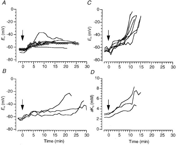
A, changes in membrane potential of DVMs induced by switching the superperfusate from one containing 3 mM K+ to one containing 10 mM KCl (−) or 10 mM K2SO4 ( ) at time zero (arrow; see Methods). B, effects on membrane potential of 2–5 μM ouabain when [K+]o was raised to 10 mM. C and D, changes in membrane potential (C) and contralaterally measured extracellular K+ activity (D) induced by 25 μM ouabain.