
Figure 11. Distributions of neurones relative to the surface of the pericruciate cortex.
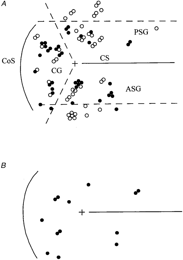
A shows locations of 46 neurones unresponsive to nerve stimulation in the resting animal (^) and 37 neurones responsive to one or both peripheral nerves (•). Upper and lower horizontal dashed lines are, respectively, 2 mm caudal and rostral to the landmark (+) provided by the lateral end of the cruciate sulcus (CS). Diagonal dashed lines approximate to the junctions between the anterior sigmoid, posterior sigmoid and coronal gyri (ASG, PSG, CG). CoS, coronal sulcus. Results from all five animals are pooled (see Methods and text). B shows the distribution of the 13 neurones for which step phase dependency could be studied (•).